Which core material is best for medical imaging tables: PMI foam or honeycomb? Closed-cell polymethacrylimide (PMI) foam delivers uniform radiolucency, isotropic shear strength, and built-in fluid resistance, while aramid honeycomb provides directional stiffness for long, flat spans. The optimal selection depends on your specific cantilever deflection limits, image artifact tolerances, and clinical disinfection requirements.
What Is the Difference Between PMI Foam and Honeycomb for Medical Imaging Tables?
PMI foam core: a closed-cell structural material
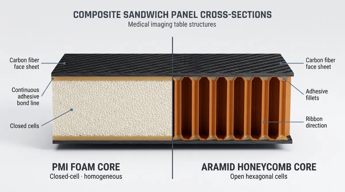
Polymethacrylimide (PMI) foam, available in high-performance grades such as ROHACELL® IG-F, is a rigid, closed-cell polymer engineered for sandwich construction. Its microstructure consists of a uniform, isotropic network of closed polyhedral cells with no internal voids or hollow channels. Because these cells are sealed, PMI foam exhibits balanced shear, tensile, and compressive performance in all spatial directions.
In medical composite manufacturing, PMI foam technology provides high thermal stability, with heat distortion resistance ranging from 190°C to 230°C depending on the specific grade. It withstands autoclave processing and compression molding cycles up to 180°C and 0.7 MPa without cell collapse. The closed-cell structure ensures that resin uptake during resin transfer molding (RTM), infusion, or prepreg processing remains confined strictly to the cut surface cells. This prevents excess resin absorption from adding parasitic weight or creating irregular, radiopaque density gradients.
Honeycomb core: a lightweight cellular structure
Aramid honeycomb consists of calendered meta-aramid paper coated with heat-resistant phenolic resin, configured into an open-cell, anisotropic layout. Developed primarily for aerospace structures, it uses hexagonal or over-expanded cells to achieve a high strength-to-weight ratio along the vertical cell axis. Most of a honeycomb core volume is air surrounded by thin composite walls.
This open architecture creates distinct directional properties. Compressive stiffness is high perpendicular to the table surface, but in-plane shear properties are anisotropic. The ribbon direction (L-direction), which follows the continuous bonded paper seam, delivers substantially higher shear strength and modulus than the transverse direction (W-direction). Structural engineers must align the ribbon axis carefully with the primary load path to avoid shear failure across the weaker orientation.
How sandwich panel construction supports imaging tables
Neither PMI foam nor honeycomb functions as a standalone table. Both act as the shear-carrying core within a composite sandwich structure. Structurally, a sandwich panel operates like an I-beam. Thin, rigid composite face sheets-typically carbon fiber, aramid, or fiberglass in an epoxy matrix-act as the flanges that carry axial tension and compression from patient weight. The structural foam core or honeycomb core serves as the web, maintaining skin separation, maximizing the second moment of area, and carrying transverse shear loads.
For medical imaging applications, the engineering goal is to maximize flexural rigidity ($D = E \cdot I$) while minimizing thickness, overall weight, and X-ray beam attenuation. Increasing core thickness increases flexural stiffness exponentially with only a linear increase in core weight. However, gantry clearances and patient transfer ergonomics impose hard physical limits on table thickness. The chosen core material must transfer shear efficiently between the face sheets while maintaining structural integrity under dynamic and localized point loads.
How Do PMI Foam and Honeycomb Compare for X-Ray and CT Imaging?
X-ray attenuation and artifact risks
In planar radiography and fluoroscopy, uniform radiolucency is just as critical as low baseline attenuation. A patient tabletop can display a low overall aluminum equivalence (mm Al) and still fail clinical requirements if localized density fluctuates across the imaging field. Abrupt local density variations show up on digital flat-panel detectors, obscuring fine bone micro-architecture or creating false indications of pathology.
PMI foam avoids this risk because its fine, closed-cell geometry creates a uniform density field. With cell diameters measured in microns, the X-ray beam traverses a statistically homogeneous cross-section, yielding an artifact-free background. In contrast, aramid honeycomb consists of alternating solid phenolic nodes, single-thickness cell walls, adhesive fillets, and hollow air chambers. During low-kVp diagnostic procedures or high-contrast fluoroscopy, this regular geometric grid can project visible pattern artifacts, phase shifts, or detector aliasing that require corrective post-processing software.
CT compatibility and image uniformity
Computed tomography amplifies core uniformity challenges because the imaging system reconstructs volumetric slices from hundreds of angular projections. Diagnostic CT tabletops reside inside the reconstruction field of view during helical acquisitions, scout scans, and pediatric examinations. Density variations within the scanning circle disrupt Hounsfield Unit (HU) calibration and create image noise.
PMI foam maintains an isotropic, uniform density that provides a flat, predictable HU baseline suitable for automated software subtraction. Honeycomb cores present variable path lengths through phenolic walls and air cavities as the X-ray tube rotates around the gantry. This angular shift in attenuation produces ring artifacts, directional streak shadows, and baseline drift beneath the patient, complicating automated image segmentation and quantitative CT analysis.
MRI compatibility and electromagnetic considerations
Material evaluation for magnetic resonance imaging centers on magnetic susceptibility, dielectric properties, and radiofrequency (RF) transparency. Unreinforced PMI foam and aramid honeycomb are both non-metallic and non-conductive, making the bare cores MRI-safe. However, full sandwich panel behavior depends on the skin and bonding configuration.
While carbon-fiber face sheets offer excellent strength, their electrical conductivity allows gradient switching and RF excitation fields to induce circular eddy currents. These currents disrupt $B_0$ magnetic field homogeneity, attenuate the RF signal, lower the signal-to-noise ratio (SNR), and risk patient heating. For MR-compatible and multi-modality designs, engineers often substitute carbon fiber with high-strength glass or aramid skins paired with a PMI foam core. Clinical acceptance standards typically require less than 10% SNR loss in the body scout field, requiring core and adhesive systems free of metallic contaminants or polar additives.
Ultrasound table requirements
Ultrasound procedure tables require clear acoustic pathways and ergonomic transducer access. Standard composite sandwich construction reflects high-frequency ultrasound waves because of acoustic impedance mismatches between human tissue, carbon skins, and cellular core materials, preventing direct trans-table scanning.
Consequently, ultrasound tables incorporate physical drop-down sections, transducer cutouts, and specialized perimeter chamfers. PMI foam delivers significant manufacturing efficiency here: it can be CNC-machined into smooth steps, drop-offs, and custom contours without compromising perimeter strength. Cutting through a honeycomb panel exposes hollow open cells that require structural edge dams, manual potting, and localized skin reinforcements to maintain stiffness.
How core thickness and skins affect radiolucency
The total radiolucency of an imaging tabletop is the sum of every material layer across the cross-section: upper skin, adhesive bond lines, core material, lower skin, and exterior surface coatings. Attenuation is quantified as aluminum equivalence at a specific beam quality (typically 100 kVp):
$$\text{Total Al-Eq} = \text{Al-Eq}_{\text{skins}} + \text{Al-Eq}_{\text{core}} + \text{Al-Eq}_{\text{adhesives}}$$
Because structural cores have a low physical density relative to composite skins, the face sheets contribute 60% to 80% of total panel attenuation despite their thin profile. A thicker core allows engineers to reduce face-sheet thickness while maintaining panel bending stiffness. However, when using honeycomb, the heavy adhesive films or foaming adhesives required to build structural fillets along thin cell walls add parasitic radiopacity, offsetting the theoretical mass advantages of the open core.
Which Core Provides Greater Strength and Table Stability?
Compressive strength under patient and equipment loads
Imaging tabletops must withstand concentrated compression from bariatric patients and heavy localized forces during emergency CPR procedures. Structural foams and cellular cores respond differently to these localized compressive stresses.
| Core Material & Grade | Nominal Density | Compressive Strength (Stabilized) | Compressive Modulus | Shear Strength (L / W) | Shear Modulus (L / W) |
|---|---|---|---|---|---|
| ROHACELL® 31 IG-F | 32 kg/m³ | 0.4 MPa | 17 MPa | 0.4 MPa (Isotropic) | 13 MPa (Isotropic) |
| ROHACELL® 51 IG-F | 52 kg/m³ | 0.9 MPa | 43 MPa | 0.8 MPa (Isotropic) | 19 MPa (Isotropic) |
| ROHACELL® 71 IG-F | 75 kg/m³ | 1.5 MPa | 73 MPa | 1.3 MPa (Isotropic) | 29 MPa (Isotropic) |
| ROHACELL® 110 IG-F | 110 kg/m³ | 3.0 MPa | 120 MPa | 2.4 MPa (Isotropic) | 50 MPa (Isotropic) |
| Aramid Honeycomb (HRH-10, 3.0 pcf) | 48 kg/m³ | 2.07 MPa | 138 MPa | 1.21 MPa / 0.69 MPa | 41 MPa / 24 MPa |
| Aramid Honeycomb (HRH-10, 4.0 pcf) | 64 kg/m³ | 3.69 MPa | 193 MPa | 1.76 MPa / 0.97 MPa | 59 MPa / 32 MPa |
Stabilized aramid honeycomb shows higher bare out-of-plane compressive strength than comparable density foam. However, honeycomb performance relies on strict perpendicular wall stabilization; off-axis compressive loads can trigger buckling of the thin walls. PMI foam provides a progressive, ductile compressive response, absorbing mechanical energy without sudden cell wall collapse.
Bending stiffness, span length, and deflection
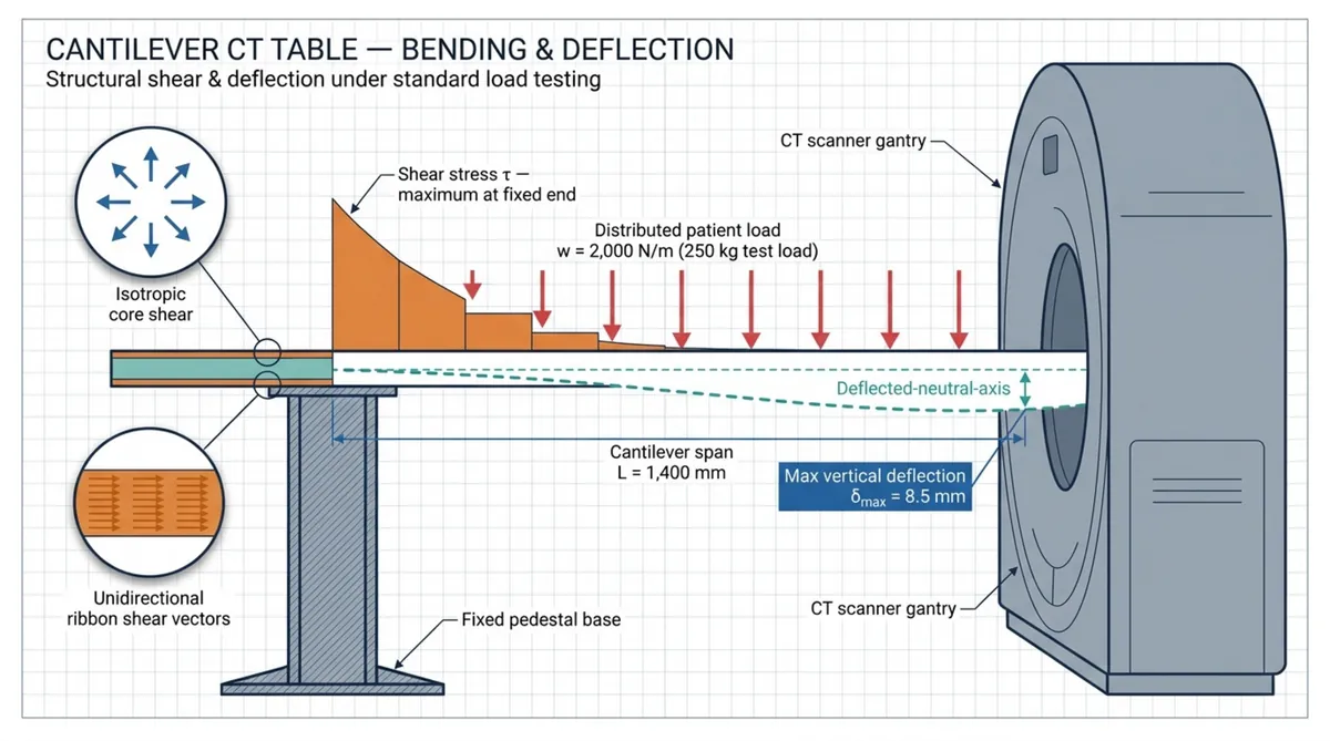
Medical table standards impose strict limits on structural deflection. Under IEC 60601-2-44, a CT patient cradle must pass rigorous vertical deflection tests under a distributed 135 kg load across a 1.9-meter cantilever span, stepped through the gantry and held to verify creep resistance. In radiation therapy, positioning limits are even tighter: vertical deviation must remain within ±0.5°, horizontal travel perpendicularity within ±1°, and absolute sagittal deflection below 2.0 mm across full travel.
Controlling deflection over a two-meter cantilever requires high structural rigidity. Aramid honeycomb provides high bending stiffness along a single axis when its ribbon (L-direction) is aligned with the longitudinal span. However, when complex load cases introduce torsional twisting or multi-axis bending-such as in tilting C-arm angiography cradles-honeycomb is limited by its lower transverse shear modulus (24 to 32 MPa). In these multi-directional load paths, the isotropic shear modulus of high-density PMI foam (29 to 50 MPa) provides superior torsional resistance and dimensional stability.
Resistance to localized loads and indentation
Patient tables frequently experience localized point loads from clinician elbows, hard transfer boards, and dropped accessories. Resistance to permanent surface indentation depends on how the core supports the outer composite skin.
Drop-impact research demonstrates that when impact energy buckles the thin walls of an aramid honeycomb core, it creates an internal void beneath the carbon skin. While the skin may spring back elastically, the core below remains crushed, leaving the laminate unsupported against subsequent fatigue loads. PMI foam spreads point loads through its adjacent closed-cell structure. This uniform backing limits core collapse, reduces micro-cracking in the face sheet, and minimizes the risk of progressive skin delamination.
Fatigue performance during repeated patient transfers
In high-throughput clinical centers, an imaging table experiences thousands of cyclic loads annually. These include dynamic patient transfers from hospital beds and cyclic cantilever deflection during motorized gantry movements.
In honeycomb construction, the adhesive fillet linking the skin to the narrow cell wall edge represents the primary fatigue bottleneck. Repeated shear reversals can generate micro-fissures in these adhesive joints, leading to skin-to-core debonding over time. PMI foam provides a continuous bonding surface across the entire composite interface. This broad contact area lowers average bond-line shear stress, delivering robust fatigue performance across millions of loading cycles.
Load ratings for fixed, mobile, and cantilevered tables
Modern clinical requirements demand higher patient weight capacities. Current regulatory clearances specify dynamic working loads up to 300 kg or 307 kg, including CPR safety margins. Fixed, mobile, and cantilevered systems support these requirements through distinct core strategies:
- Cantilevered CT and Angiography Tables: Supported only at the pedestal root, these tables carry severe bending and shear stresses near the mounting zone. Engineers often use graded density cores, placing high-shear PMI foam (such as ROHACELL® 110) at the cantilever root and transitioning to lower-density cores along the patient span.
- Mobile and C-Arm Surgical Tables: These systems rely on dual supports or mobile bases where overall weight determines manual handling safety. Honeycomb allows weight savings for long, purely flat spans, helping keep mobile equipment within manual handling limits.
How Do PMI Foam and Honeycomb Perform in Clinical Environments?
Moisture absorption and fluid resistance
Clinical imaging environments present frequent exposure to physiological fluids, saline solutions, contrast agents, and liquid disinfectants. Fluid ingress into a composite sandwich panel compromises structural integrity and introduces contamination risks.
PMI foam has a sealed, closed-cell morphology that eliminates fluid migration through capillary action. If an impact punctures the outer carbon skin, fluid remains confined to the damaged surface cells. In contrast, aramid honeycomb is an open-cell network. Once the protective skin is breached, fluids enter the hollow cell chambers. Gravity, capillary action, and patient-induced pressure cycles pump liquid throughout the core. Trapped fluids cannot be sanitized, creating biological contamination risks, permanent radiopaque artifacts on diagnostic images, and long-term adhesive debonding.
Cleaning agents, disinfectants, and chemical compatibility
Hygienic protocols require thorough chemical wiping of imaging tables between patients. Standard hospital-grade disinfectants include:
- Quaternary ammonium compounds
- Sodium hypochlorite (clinical bleach solutions)
- Accelerated hydrogen peroxide (AHP)
- Isopropyl and ethyl alcohol solutions (70% to 90%)
- Phenolic surface germicides
High-integrity epoxy matrices and polyurethane clearcoats serve as the primary chemical barrier. However, aggressive sanitizers can penetrate through micro-cracks, mounting holes, and perimeter joints over years of service. PMI foam provides chemical resistance against common alcohols, aliphatic hydrocarbons, and clinical disinfectants. Aramid honeycomb exposed to cleaning solutions can suffer gradual degradation of its phenolic dipping resin from alkaline or oxidizing agents, leading to softened cell walls and reduced compressive modulus.
Temperature stability and sterilization limits
While diagnostic imaging runs at ambient clinical temperatures, thermal performance is critical during composite manufacturing and medical sterilization cycles. Medical sandwich panels must remain thermally stable under processing temperatures.
Aramid honeycomb maintains dry-heat stability up to 200°C-230°C before the phenolic matrix softens. PMI structural foam provides exceptional thermal performance under controlled composite processing, with standard ROHACELL® grades processing up to 130°C at 0.3 MPa, and high-performance grades tolerating up to 180°C and 0.7 MPa during autoclave curing. At room temperatures (18°C to 24°C), the coefficient of thermal expansion of PMI foam ($25\text{-}35 \times 10^{-6}/\text{K}$) matches epoxy resin systems well, preventing thermal print-through on thin carbon-fiber skins.
Impact damage, crushing, and hidden core failure
A major structural concern in composite tables is Barely Visible Impact Damage (BVID). This occurs when dropped items-such as heavy syringe shields, RF coils, or handheld probes-crush the core without leaving a significant visual crack on the carbon skin.
Because aramid honeycomb has ultra-thin cell walls, impact energy can buckle the core internally. The elastic face sheet springs back, concealing a hollow, unsupported pocket underneath. Subsequent patient loading subjects the unsupported skin to cyclic bending, accelerating delamination. PMI foam absorbs impact energy through controlled, uniform deformation across its cell structure, maintaining local shear support and preventing rapid damage propagation.
Service life and maintenance requirements
Hospitals expect diagnostic imaging tables to maintain structural and aesthetic performance over a 7- to 10-year service life across tens of thousands of procedures.
PMI-cored panels require less maintenance over their lifecycle because their closed cells block fluid migration and make repairs straightforward. A localized puncture can be drilled out, filled with an epoxy-microballoon compound or matched foam plug, and capped with a flush composite ply patch. In contrast, repairing an open honeycomb core contaminated with fluids requires vacuum moisture extraction, extensive core excision, and dense potting, which introduces localized radiopaque artifacts into the imaging area.
What Are the Manufacturing and Design Trade-Offs?
Machining complex shapes and integrated features
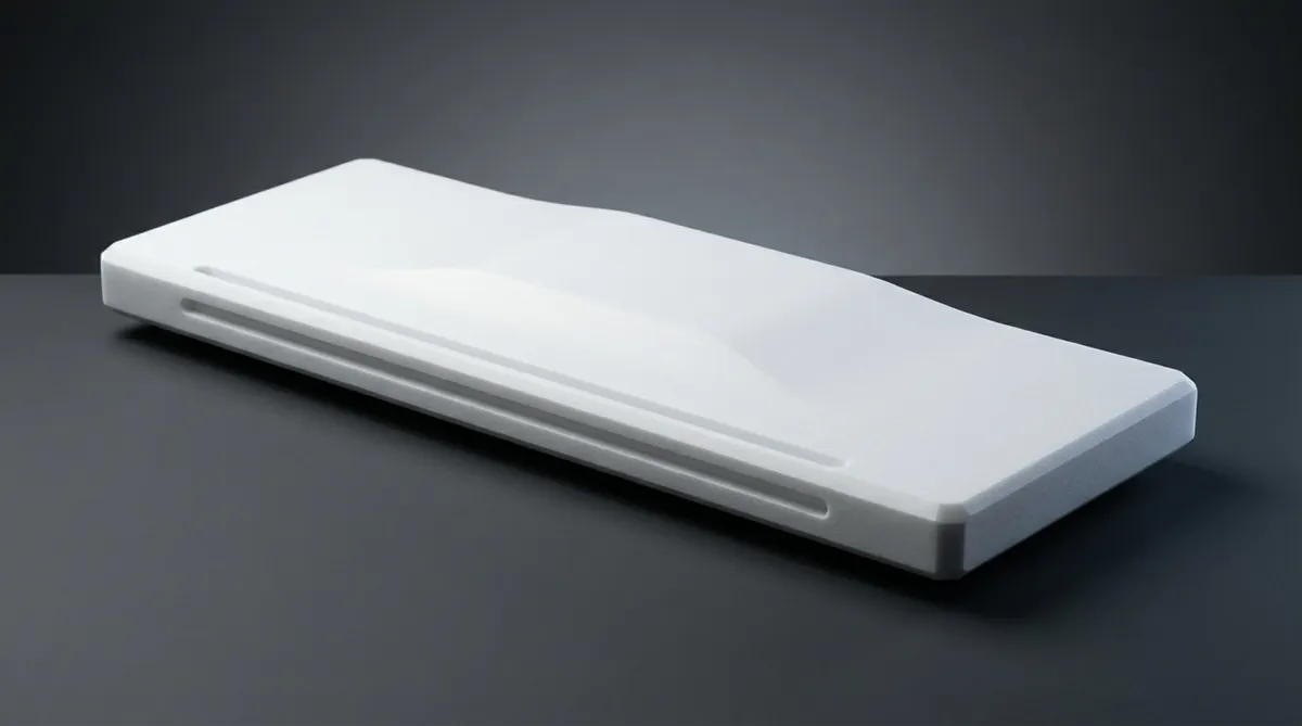
Modern imaging tabletops require complex contours, including contoured head sections, edge chamfers for patient transfers, gantry clearances, and recessed accessory channels.
PMI foam offers exceptional machinability. It can be milled, routed, turned, and drilled to sub-millimeter tolerances using standard high-speed cutting tools without lubricants. Chem-Craft provides tight-tolerance foam shape milling directly from customer CAD models, enabling complex 3D profiles, tapers, and pocketed geometries from a single foam blank. Honeycomb is significantly more difficult to machine cleanly: high-speed milling can tear, burr, or crush unsupported cell walls, requiring specialized roll cutters, sacrificial fill waxes, or cryogenic freezing.
Bonding skins, inserts, rails, and mounting hardware
Imaging tabletops require robust attachment points for surgical accessory rails, drive pedestals, patient restraint straps, and cable management channels. Thin face sheets cannot support these localized point loads independently.
Each core material dictates a specific fastening approach:
- In Honeycomb Structures: Mechanical fasteners cannot thread directly into open cells. Fabricators must mill out pockets, position metal or thermoplastic bobbins, and inject dense syntactic potting compound. This multi-step process adds labor, weight, and radiopaque artifacts.
- In PMI Foam Structures: Engineers can mill precise pockets and bond inserts directly into the core. In moderately loaded zones, local inserts can be eliminated by bonding higher-density foam inserts (such as ROHACELL® 110) within a lower-density field (such as ROHACELL® 51). This approach transfers hardware loads smoothly without creating dense potting zones that show up on scans.
Core thickness, density, and panel weight
Total tabletop weight directly influences the engineering requirements of the scanner drive system. Lower mass reduces power consumption and allows smaller servo motors, lighter drive screws, and smaller counterweights. Nomex honeycomb offers an initial mass advantage, with common grades weighing 48 kg/m³ while providing high out-of-plane strength.
However, total sandwich panel mass depends on resin consumption during manufacturing. In honeycomb construction, prepregs and film adhesives form generous fillets along the vertical cell edges. Without strict process controls, these fillets add 200 to 400 g/m² of unreinforced adhesive mass per bond line. PMI foam features a fine, closed-cell surface that limits resin absorption to the open cut cells, drawing less than 100 g/m² during controlled resin infusion or prepreg bonding. Consequently, the finished weight of a PMI sandwich panel often matches that of a honeycomb structure, despite the higher nominal density of the raw foam core.
Dimensional tolerance and surface flatness
Stereotactic radiation therapy and high-resolution CT require high surface flatness and thickness consistency to ensure accurate dose delivery and image registration. Core thickness variations directly alter X-ray attenuation and patient positioning accuracy.
Commercial aramid honeycomb sheets typically hold thickness tolerances of ±0.15 mm to ±0.25 mm, with nominal density variations up to ±10%. Chem-Craft supplies structural PMI foam via high-precision horizontal cutting down to 1 mm thicknesses with tolerances within ±0.1 mm, and can achieve flat dimension tolerances down to ±0.05 mm upon request. Furthermore, the solid backing of PMI foam eliminates cell print-through and telegraphing, ensuring a Class A surface finish on thin carbon skins.
Production volume, lead time, and total cost
When selecting core materials, engineers must balance raw stock prices against total manufacturing touch-labor.
| Factor | Aramid Honeycomb Architecture | PMI Foam Architecture |
|---|---|---|
| Raw Material Cost | Moderate to High (driven by aramid paper synthesis and phenolic dipping) | High (driven by specialized polymer chemistry and thermal expansion) |
| Tooling & Setup Expense | High (requires specialized core stabilization and machining fixtures) | Moderate (standard CNC routing and conventional composite tooling) |
| Touch-Labor Requirements | High (edge potting, insert potting, cell edge stabilization) | Low to Moderate (direct 3D machining, single-step co-curing) |
| Defect & Scrap Risk | Moderate (susceptible to cell collapse, crushed edges, and telegraphing) | Low (predictable mechanical response and uniform resin bonding) |
| Edge-Closeout Requirements | Mandatory perimeter dams, high-density edge potting, or bonded channels | Self-supporting closed perimeter; direct skin wrap-around |
While raw aramid honeycomb may carry a lower initial purchase price, the extensive touch-labor required for edge potting, insert preparation, and core-fill dams increases finished-part cost. PMI foam simplifies composite manufacturing by supporting single-step co-curing and net-shape CNC milling, reducing touch-labor and scrap rates in serial production.
What Questions Should Engineers Ask Before Selecting the Core Material?
Which ASTM, ISO, IEC, or medical-device requirements apply?
Medical table design must comply with strict international regulatory standards. Before selecting a core, design teams should verify the specific requirements for the equipment class:
- IEC 60601-1 (General Safety & Essential Performance): Mandates structural safety factors typically between $2.5\times$ and $4.0\times$ working load ratings, including dynamic push-stop loads and impact tests.
- IEC 60601-2-44 (Particular Requirements for X-Ray Systems for CT): Defines cantilever deflection limits, positioning repeatability, and structural creep verification under 135 kg loads over extended hold times.
- AAPM / ESTRO Radiation Therapy Guidelines: Establish strict geometric flatness, deflection thresholds, and beam attenuation consistency to ensure stereotactic radiation targeting accuracy.
- ISO 10993 (Biocompatibility): Regulates patient-contact safety and chemical off-gassing, verifying that composite matrices and cores emit no toxic compounds.
Specifying these standards early ensures your engineering team selects a core density and thickness that meets regulatory deflection and safety limits without over-engineering panel mass.
Has the complete finished panel been tested for image artifacts?
Evaluating raw core samples alone is insufficient for artifact verification. Clinical radiolucency depends on the consolidated composite sandwich working as an integrated system under production conditions.
Artifact testing must evaluate:
- The complete carbon or fiberglass skin schedule, including resin distribution
- Structural adhesive film layers, with focus on adhesive pooling and fillet geometry
- Core chamfers, internal splices, and step-down transitions
- Perimeter closeouts, edge dams, and embedded metallic or plastic hardware
- Antimicrobial topcoats and exterior cosmetic clearcoats
Consolidated production prototypes should undergo flat-field digital radiography and volumetric CT imaging across standard diagnostic ranges (70 to 140 kVp). This testing confirms that the core and adhesive interfaces introduce no phase shifts, ring artifacts, or Hounsfield Unit anomalies within the diagnostic field.
How Should You Choose PMI Foam or Honeycomb for a Medical Imaging Table?
Choose PMI foam for moisture exposure and complex geometry
PMI foam is the preferred engineering choice when tabletops require three-dimensional contours, fluid resistance, and uniform radiolucency. Surgical C-arm cradles, interventional fluoroscopy tables, and angiography systems face constant exposure to blood, saline, and chemical disinfectants. In these applications, closed-cell PMI foam prevents fluid ingress, safeguarding structural integrity and clinical hygiene.
Furthermore, when your design includes ergonomic contours, gantry clearance tapers, or integrated accessory drop-downs, PMI foam can be machined directly to final dimensions. Its isotropic properties ensure that complex 3D profiles maintain reliable shear and compressive strength throughout the part.
Choose honeycomb for large, lightweight, high-stiffness panels
Aramid honeycomb is well suited for long, flat, single-axis cantilevered decks where the primary engineering priority is minimizing longitudinal bending deflection while reducing total tabletop weight. General radiography tables, patient transfer decks, and standard diagnostic CT tables benefit from honeycomb’s high directional strength-to-weight ratio.
When the component features uniform thickness and a dominant longitudinal bending load, aligning the honeycomb ribbon in the L-direction provides excellent flexural stiffness ($E \cdot I$). This enables thin-profile tables that meet deflection limits under bariatric patient loads while keeping driving-servo power requirements low.
Match the core to X-ray attenuation and modality requirements
Imaging modality requirements dictate permissible core structures. For high-resolution planar X-ray, mammography, and digital subtraction angiography (DSA), avoiding structural background patterns is essential. PMI foam features an ultrafine cell structure that eliminates the shadow patterns and phase artifacts associated with open honeycomb cells.
In multi-modality environments like PET/CT or MR-guided therapy, pairing PMI foam with non-conductive skins (such as high-strength glass or aramid) prevents eddy currents and radiofrequency distortion. This preserves signal-to-noise ratio and ensures image clarity across modalities.
Evaluate patient weight, safety factors, and deflection limits
With patient weight ratings routinely exceeding 300 kg, structural engineers should evaluate the core using the following systematic approach:
- Calculate Maximum Cantilever Moments: Determine maximum bending moments and shear stresses at the table root under maximum patient loads combined with CPR forces.
- Evaluate Core Shear Capacity: For multi-axis or torsional loads, specify the isotropic shear capacity of high-density PMI foam (such as ROHACELL® 71 or 110) to prevent edge failure. For simple unidirectional bending, lower-density aramid honeycomb can meet shear requirements at minimal weight.
- Verify Local Indentation Limits: Confirm that face-sheet thickness and core compressive modulus are sufficient to resist localized impact from accessory mounts, elbows, and transfer boards.
Compare lifecycle cost rather than material price alone
Purchasing evaluations should consider total manufacturing touch-labor and operational lifecycle costs rather than raw board-foot material prices. While raw aramid honeycomb often has a lower initial material cost, it requires extensive labor for perimeter sealing, potted insert installation, and cell-stabilization steps.
PMI foam reduces manufacturing labor by supporting single-step co-curing, net-shape CNC milling, and self-supporting perimeter geometry. In clinical service, its sealed closed-cell structure prevents fluid contamination and simplifies repairs, delivering lower total lifecycle costs for hospitals and equipment manufacturers.
Verify performance through load, fatigue, cleaning, and imaging tests
Engineers should validate final core selection through a comprehensive qualification program using production-representative composite panels:
- Creep and Deflection Testing: Apply full working loads (up to 300 kg) across the cantilever span for extended periods to confirm compliance with IEC 60601-2-44 deflection and alignment limits.
- Dynamic Cyclic Fatigue: Subject the table to tens of thousands of translation, elevation, and side-transfer cycles to verify long-term core-to-skin bond integrity.
- Chemical Exposure Aging: Expose perimeter joints, fastener penetrations, and wear surfaces to hospital disinfectants to verify chemical resistance and ensure no fluid ingress.
- Full-Field Diagnostic Imaging Audits: Scan the consolidated tabletop across diagnostic energies (70 to 140 kVp) to ensure an artifact-free background and verify Hounsfield Unit consistency.
Advanced diagnostic systems such as photon-counting CT (PCCT) and AI-driven image reconstruction are increasing sensitivity to subtle core density variations. Small cell inconsistencies, resin pooling, and core defects that were smoothed by older detectors can now appear as clinical artifacts. If you need assistance selecting or machining the right structural core for your medical composite application, contact Chem-Craft to discuss your technical specifications with our engineering team.