How does PMI foam improve the performance of mammography plates and medical equipment housings? Polymethacrylimide (PMI) structural foam cores deliver high stiffness-to-weight ratios alongside minimal X-ray attenuation, providing rigid structural support without compromising diagnostic image clarity. In composite sandwich construction, this closed-cell material meets demanding engineering requirements for radiolucency, dimensional stability, and low weight across clinical environments.
What Is PMI Foam for Mammography Plates and Medical Housings?
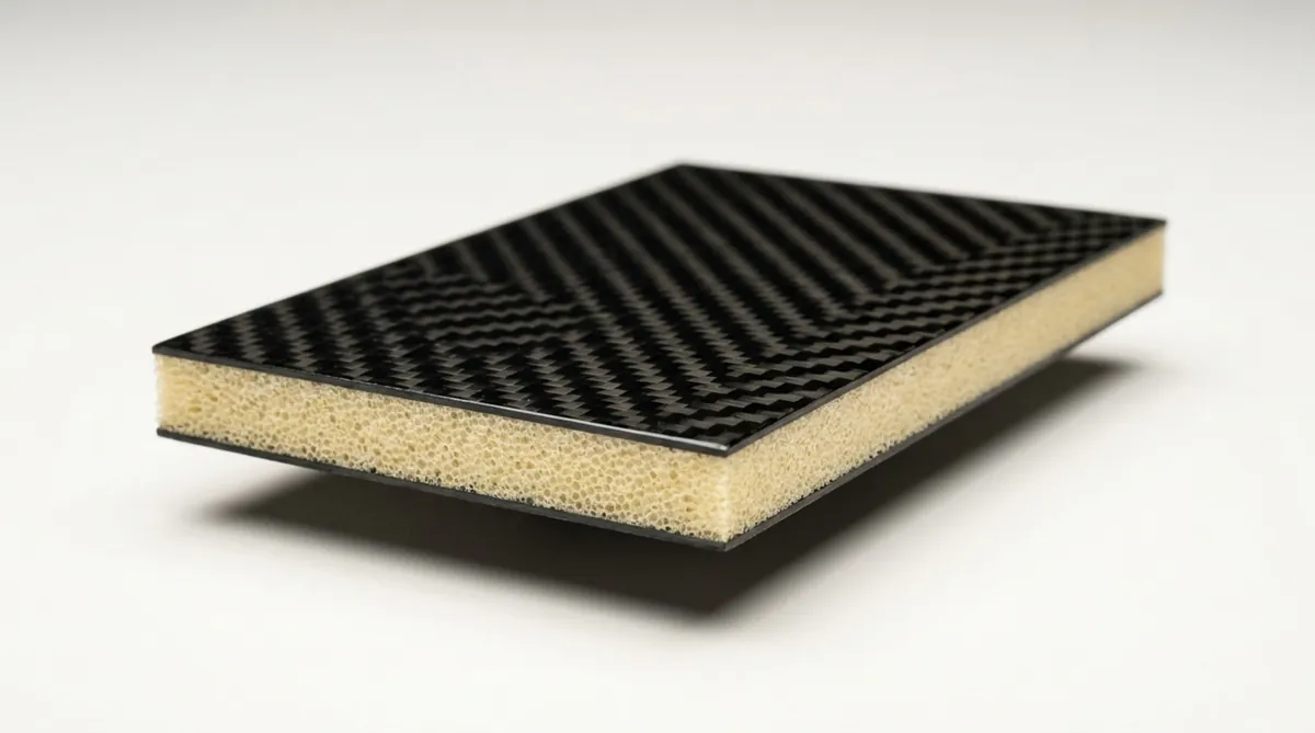
Polymethacrylimide (PMI) foam is a high-performance rigid cellular core material engineered for structural sandwich panels. In medical diagnostic equipment, engineers specify PMI when a component must handle static and dynamic mechanical loads without attenuating ionizing radiation or adding unnecessary mass.
Using PMI foam cores in mammography support plates and equipment housings allows engineering teams to balance structural integrity with radiolucency, avoiding the performance trade-offs common to legacy materials.
PMI foam composition and closed-cell structure
PMI foam is manufactured by expanding a copolymer sheet consisting primarily of methacrylic acid and methacrylonitrile. During the foaming process, heat activates an imidization reaction, creating a rigid, highly cross-linked polyheterocyclic ring system. This molecular network gives PMI higher mechanical strength and thermal stability than standard linear polymers.
The resulting material exhibits a 100% closed-cell structure with uniform, isotropic, polyhedral cell geometry. Because the cells are completely sealed, fluids cannot penetrate through capillary action. The foam repels moisture, humidity, and liquid adhesives during processing. This consistent cell structure provides predictable, multi-directional mechanical properties across every sheet.
Why polymethacrylimide foam suits medical imaging equipment
Components located directly within an X-ray beam must interact with as few photons as possible. PMI foam consists almost entirely of low-atomic-number elements: carbon, hydrogen, nitrogen, and oxygen. It exhibits an exceptionally low linear attenuation coefficient, making it virtually transparent to flat-panel digital radiography detectors.
Diagnostic equipment also requires strict mechanical stability. Mammography devices use localized compression loads to position patient tissue, requiring repeatable dimensional accuracy across thousands of scans. PMI foam technology features a rigid molecular architecture that resists compressive creep and long-term deformation under sustained clamping forces.
PMI foam core versus solid plastic and metal components
Compared to structural metals like aluminum or magnesium, PMI foam cores significantly reduce component weight while eliminating secondary radiation scatter and ghosting artifacts. Solid metal components in the imaging path cause beam hardening and scattering, reducing image contrast when detecting tiny microcalcifications.
Compared to solid engineering plastics such as polycarbonate, PEEK, or ABS, a PMI composite sandwich panel yields significantly higher flexural rigidity at a lower overall mass. Thin solid plastics bend under lateral mechanical loads, often requiring reinforcing ribs that project visible density variations onto digital images. A PMI structural foam core delivers continuous support across the entire surface without internal ribs.
What Medical Equipment Uses PMI Foam Components?
PMI foam is primarily specified in diagnostic and therapeutic devices where mechanical accuracy, lightweight structural performance, and high radiolucency are mandatory engineering requirements. Applications span routine screening instruments to advanced radiotherapy systems.
Modern clinical facilities demand equipment that supports ergonomic handling, rapid patient positioning, and high diagnostic resolution. Consequently, sandwich structures utilizing PMI foam have replaced conventional cast metals and dense thermoplastics in critical medical subassemblies.
Mammography plates, detector supports, and compression assemblies
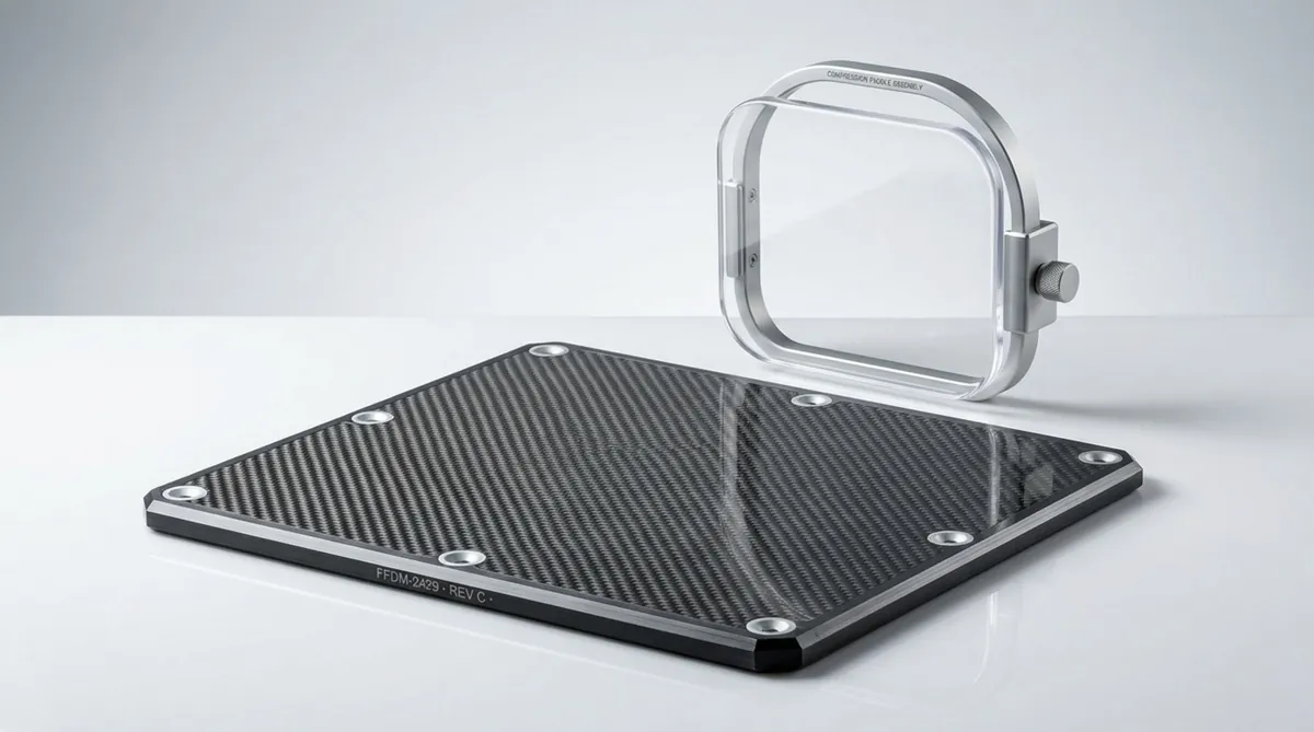
In full-field digital mammography and digital breast tomosynthesis (DBT), PMI foam serves as the central core for patient support tables, breast platforms, and flat-panel detector covers. These assemblies must remain flat through continuous examination cycles while permitting low-energy X-rays (typically 25 to 35 kVp) to reach amorphous selenium or cesium iodide detectors without beam disruption.
In biopsy attachments and compression mechanisms, PMI cores preserve component parallelism. When motorized actuators apply clamping loads, the sandwich panel resists deflection, preserving focal-plane alignment and preventing image blur that could obscure early-stage calcifications.
X-ray, CT, and other radiolucent imaging housings
Computed tomography (CT) scanners rely on PMI foam cores for patient cradles and table extensions. A CT patient table acts as an extended cantilever beam, supporting patient loads exceeding 150 to 200 kg while indexing smoothly through the gantry. Carbon-fiber composite skins bonded to a high-performance PMI core provide necessary bending resistance without creating ring artifacts or absorbing excessive radiation.
Mobile C-arm assemblies, angiography tables, and portable digital radiography cassettes also utilize PMI cores. In these composite applications, the material minimizes secondary radiation scatter and supports high Modulation Transfer Function (MTF) performance, helping clinical teams maintain lower exposure parameters.
Protective housings for medical electronics
Beyond the direct X-ray beam, digital signal-processing electronics require protection against mechanical shock, operational vibration, and thermal fluctuations. Handheld ultrasound scanners, portable digital flat-panel detectors, and mobile surgical robotics utilize PMI sandwich enclosures to achieve durable, impact-resistant protection.
These composite shells protect sensitive analog-to-digital converters and solid-state sensors from impact damage during daily transport. Furthermore, PMI foam exhibits low thermal conductivity (approximately 0.030 to 0.035 W/m·K), effectively shielding delicate sensor arrays from heat generated by internal power-management components, thereby suppressing thermal noise in diagnostic data.
Which PMI Foam Properties Matter for Mammography and Medical Devices?
Selecting an appropriate foam grade requires evaluating imaging transparency, structural capabilities, and manufacturing process compatibility. A core that meets static strength metrics may still cause imaging artifacts if the cellular geometry is coarse or irregular.
Design teams must assess how microscopic foam characteristics dictate macro-level performance under continuous clinical usage.
Density, compressive strength, and elastic modulus
Structural PMI foams, such as Rohacell IG-F and Rohacell HF, are produced in densities ranging from 30 kg/m³ to over 200 kg/m³. Medical sandwich structures typically incorporate low- to medium-density grades, including 32, 52, and 75 kg/m³. Compressive strength scales directly with density, ranging from approximately 0.4 MPa in ultra-light grades to well over 1.5 MPa in medium-density variants.
Elastic modulus follows an identical trend, providing the core with the shear modulus required to prevent core shear failure during flexure. Engineering teams select the core density based on peak localized loads near mounting brackets, balancing necessary structural integrity against the superior radiolucency of lower-density grades.
Cell size, thickness, and core uniformity
Cell size is critical for materials placed directly in the radiation beam. Fine, consistent cell diameters-typically between 0.3 mm and 0.6 mm, characteristic of grades such as Rohacell RIMA-prevent granular background patterns from appearing on high-resolution digital scans. Larger or uneven cell structures can create subtle density gradients on the sensor, complicating the detection of fine tissue features.
Tight thickness tolerances are equally important. Variations in sheet thickness cause uneven resin pooling during sandwich consolidation. A thickness variation of just 0.1 mm can create a local resin pocket during vacuum processing, introducing an area of elevated X-ray attenuation that mimics biological pathology.
Radiolucency and imaging-path compatibility
PMI radiolucency depends on elemental mass distribution within the X-ray trajectory. Laboratory data generated with 100 kV diagnostic spectra confirms that a 2 mm section of low-density PMI transmits over 99.5% of incident radiation. Even thick sections reaching 45 mm maintain transmission levels above 95.9%, ensuring diagnostic photon losses remain negligible.
Uniformity of attenuation is as vital as net transmission. Structural foam cores must be entirely free from heavy-element contaminants, voids, and resin inclusions. Medical-grade PMI cores undergo controlled production to ensure foreign elements with higher atomic numbers, such as iron or zinc, do not contaminate the foam matrix.
Temperature resistance and dimensional tolerances
PMI foams exhibit high heat distortion resistance. Specific grades withstand composite cure cycles up to 180°C or even 200°C without cell collapse under pressure, making them suitable for aerospace-grade carbon-fiber prepreg systems cured in autoclaves or heated presses.
Precise dimensional stability ensures finished sandwich panels align with fine-pitch digital detectors. PMI foam has a low coefficient of thermal expansion (CTE) that closely matches carbon-fiber reinforced polymers (CFRP), ensuring assemblies remain flat despite temperature shifts between active internal electronics and climate-controlled radiology suites.
Flammability, smoke, and outgassing requirements
Medical devices operating in clinical environments must comply with fire safety requirements, including UL 94 standards. PMI foam is inherently self-extinguishing due to its chemical cross-linking, though flammability characteristics depend on core density and structural formulation.
Specific flame-resistant grades satisfy UL 94 V-0 requirements, minimizing flame spread and smoke emissions. Furthermore, low outgassing characteristics prevent chemical vapors from condensing on sensitive optical mirrors, solid-state detector arrays, or internal electronics during prolonged operation.
How PMI Foam Supports Mammography Plate Performance
Mammography demands exceptional spatial resolution and contrast sensitivity. Because the optical contrast between normal glandular tissue and microcalcifications is minute, every component within the beam path must be engineered with precision.
Integrating a structural PMI foam core into breast support tables provides the mechanical stiffness required by clinical standards while maximizing X-ray throughput.
Low X-ray attenuation for clearer image transmission
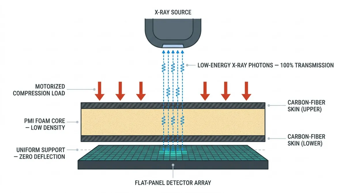
Full-field digital mammography utilizes low tube potentials, typically between 25 and 32 kVp. Photons generated in this energy spectrum are readily absorbed or scattered by dense structural materials. PMI foam consists of light elements that minimize both photoelectric absorption and Compton scattering, allowing the maximum number of primary photons to strike the detector.
Minimizing attenuation preserves image contrast, which is essential for identifying calcifications smaller than 0.2 mm. It also aids compliance with the ALARA (As Low As Reasonably Achievable) principle, helping keep the average glandular dose (AGD) well below the regulatory threshold of 3.0 mGy per craniocaudal exposure specified by the Mammography Quality Standards Act (MQSA).
High stiffness-to-weight ratio for stable plate support
During patient positioning, mammography systems apply steady motorized compression to flatten breast tissue, separate overlapping anatomical features, and reduce motion blur. Standard protocols mandate motorized compression loads between 111 N and 200 N, requiring the compression support structure to remain parallel with less than 1.0 cm of deflection across the panel.
A PMI sandwich panel satisfies these deflection limits with very low structural mass. By separating the load-bearing CFRP face skins, the foam core substantially increases the panel’s area moment of inertia, resisting flexure and maintaining uniform tissue thickness throughout the exposure.
Dimensional stability during repeated imaging cycles
A high-throughput screening facility may execute dozens of examinations daily, subjecting the patient support platform to cyclical compression and release. Lower-grade cellular materials can suffer fatigue degradation, cell-wall fracture, or permanent creep deformation, resulting in panel sagging over time.
PMI foam provides excellent compressive creep resistance and cyclic fatigue endurance. The imidized cell structure recovers elastically following compression cycles, maintaining surface flatness within fractions of a millimeter through hundreds of thousands of imaging procedures. This geometric stability ensures reliable spatial calibration for digital breast tomosynthesis reconstruction algorithms.
Lightweight construction that improves equipment handling
Ergonomics play a direct role in clinical examination efficiency. Technologists continuously reposition, elevate, and tilt the mammography gantry between craniocaudal (CC) and mediolateral oblique (MLO) orientations. Bulky, heavy assemblies impede rapid adjustments and contribute to operator strain.
Replacing solid plastic or metallic plates with optimized PMI sandwich structures dramatically lowers moving gantry mass. This weight reduction reduces wear on drive motors, simplifies counterweight mechanisms, and enables smoother equipment manipulation during daily screening operations.
What Benefits Does PMI Foam Provide in Medical Housings?
Medical enclosures protect optoelectronics, high-voltage assemblies, and digital acquisition sensors from impacts, fluid exposure, and mechanical vibration. They also establish the physical architecture and ergonomic feel of the equipment.
Employing PMI structural foam cores within composite housing shells offers engineering benefits that conventional thermoformed plastics cannot match.
Reduces housing weight without sacrificing rigidity
Point-of-care ultrasound carts, mobile C-arms, and portable bedside radiography units require chassis covers that offer high rigidity at minimal weight. A thin sandwich shell containing a PMI foam core achieves higher flexural stiffness than thick monolithic polyurethane or stamped aluminum sheets.
This structural efficiency enables healthcare personnel to maneuver heavy diagnostic systems through tight clinic rooms and elevator doors with less physical effort. The core eliminates panel flexing, providing a rigid feel without adding dead weight.
Improves impact resistance and vibration control
Mobile medical carts routinely encounter impacts with hospital beds, door jambs, and structural walls. The uniform closed-cell matrix of PMI foam absorbs kinetic energy through elastic deformation of cell struts, dispersing point-load stresses across the sandwich panel to protect internal subsystems.
The cellular matrix also provides mechanical damping against operational vibration. In diagnostic assemblies equipped with cooling blowers, motorized gantries, or rotating X-ray tube anodes, PMI housing panels suppress high-frequency acoustic and structural resonance, creating a quieter environment for patients.
Supports thermal insulation around sensitive electronics
Diagnostic equipment frequently places heat-generating power supplies adjacent to imaging sensors that are highly sensitive to thermal gradients. CMOS and amorphous silicon flat-panel detectors exhibit thermal drift when subjected to localized heating, increasing electronic noise and causing calibration shifts.
PMI foam serves as an effective thermal insulator with a thermal conductivity rating around 0.030 to 0.035 W/(m·K). Incorporating the foam into internal partitions and external housings thermally isolates detector arrays from adjacent heat sources, maintaining stable sensor operating temperatures and reducing the frequency of dark-field recalibrations.
Resists moisture absorption and chemical exposure
Hospital staff clean medical hardware daily using aggressive chemical agents, including quaternary ammonium compounds, isopropyl alcohol, and sodium hypochlorite solutions. Unreinforced solid thermoplastics can develop environmental stress cracking (ESC) when exposed to these chemicals under residual molding stress.
PMI foam demonstrates broad chemical stability against typical clinical disinfectants. Its closed-cell geometry prevents fluid ingress, protecting the core from microbial growth or fluid-induced degradation, provided panel perimeters and penetrations are correctly sealed with suitable barrier resins.
Enables thin-wall and sandwich-panel housing designs
Conventional structural plastics require extensive networks of internal support ribs, bosses, and gussets to prevent large surfaces from buckling. These features occupy internal volume and often cause visual sink marks on exterior cosmetic surfaces.
PMI cores allow engineers to manufacture large, self-supporting sandwich panels with completely smooth profiles. This eliminates internal ribs, maximizing usable volume for cabling, cooling channels, and electronics, while facilitating easy cleaning on outer surfaces.
How Is PMI Foam Integrated into Mammography Plates and Medical Housings?
Transforming raw PMI foam into finished medical components requires established composite manufacturing methods. These workflows involve precision CNC cutting, composite lamination, and controlled curing schedules to preserve cell structure integrity.
Composite engineers must manage each production step to prevent dry spots, localized crushing, or resin pooling that could compromise structural metrics or create imaging artifacts.
CNC machining, cutting, and contour forming
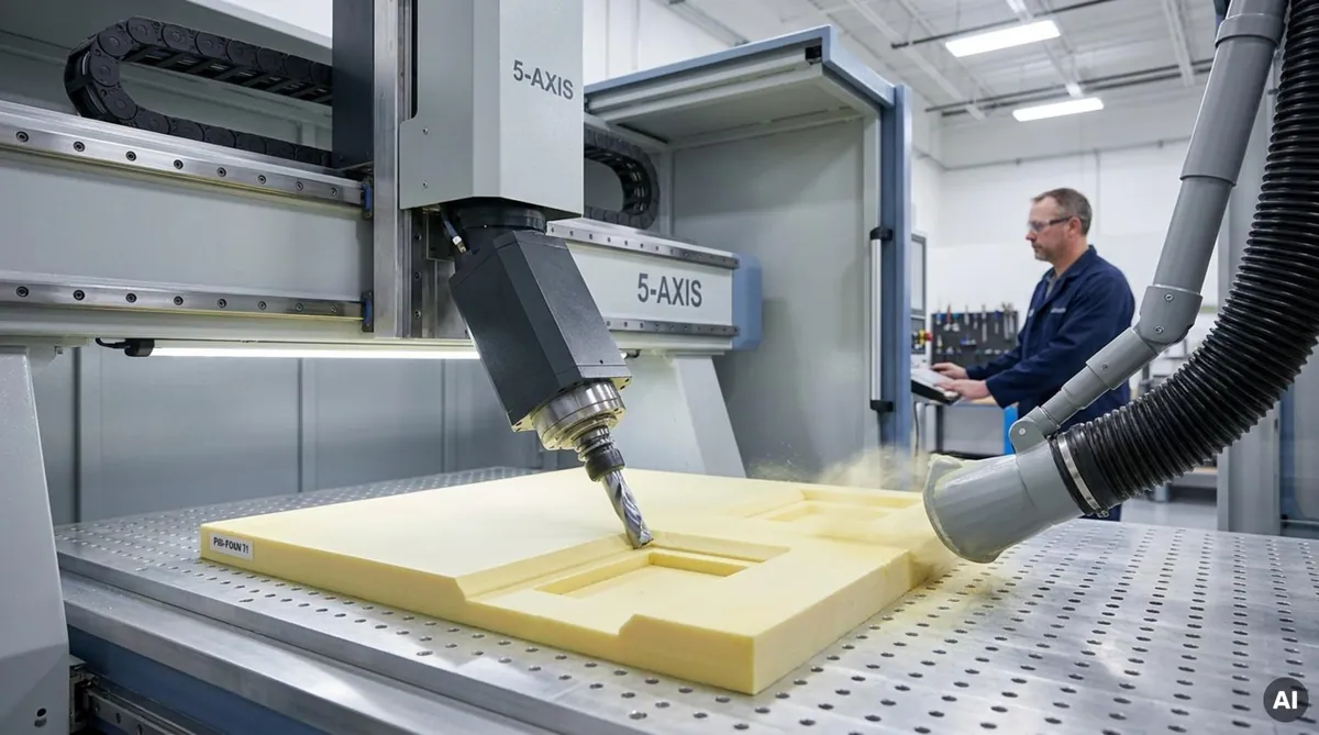
PMI foam is manufactured in large blocks and subsequently sliced into calibrated sheets. At Chem-Craft, horizontal cutting services supply foam sheets in thicknesses from 1 mm up to maximum block dimensions, holding tight thickness tolerances down to 0.1 mm. Machining intricate shapes and edge reliefs is accomplished using multi-axis CNC milling equipment. Because PMI behaves as a rigid-elastic solid, sharp carbide tooling cuts the foam cleanly without tearing cells or creating surface fuzz.
For curved components such as gantry fairings, PMI can be thermoformed at temperatures between 165°C and 205°C. When heated uniformly, the polymer softens into an elastic state, allowing it to be formed over single- or compound-curvature tooling using vacuum pressure or matched molds. Upon cooling below 100°C, the core permanently sets in the target geometry with minimal spring-back.
Bonding PMI foam to carbon fiber, composites, or thermoplastics
Sandwich panel performance depends on the shear transfer between the core and face sheets. Mammography plates typically utilize intermediate- or high-modulus carbon-fiber fabrics impregnated with toughened epoxy matrices.
During composite lay-up, the PMI core is positioned between prepreg layers. Due to its high compressive creep resistance, the assembly can undergo co-curing under vacuum or inside an autoclave at temperatures up to 180°C and pressures up to 0.7 MPa. Prepreg resin flows slightly into the surface cells of the core, creating a strong mechanical interlock without requiring heavy auxiliary adhesive films.
Using PMI foam in lightweight sandwich panels
Designing composite panels requires calculating core shear limits alongside tensile and compressive limits for the face sheets. In a cantilevered mammography breast platform, the CFRP face skins carry the longitudinal bending loads, while the PMI core transfers shear forces and prevents skin wrinkling or buckling under compression.
Engineers often design stepped-density cores to optimize performance. In the central X-ray aperture, an ultra-low-density foam core (such as 32 or 52 kg/m³) provides maximum radiolucency. Around the perimeter and mechanical mounting points, higher-density foam grades (such as 75 or 110 kg/m³) are spliced into the panel to carry elevated localized shear loads.
Adding skins, inserts, fasteners, and mounting features
Low-density structural foams cannot support high localized pull-out or clamping forces. Fasteners must never be threaded directly into raw PMI material. Instead, sandwich assemblies incorporate engineered hardpoints for mechanical attachment.
Engineers position potted metallic inserts, solid composite perimeter blocks, or aluminum hardpoints into the core before lamination. Structural epoxy potting compounds bond these inserts securely, distributing mechanical loads from hinges, actuators, and mounting rails evenly across the sandwich core.
Controlling tolerances around detectors and electronic assemblies
Modern flat-panel digital detectors operate with pixel pitches between 50 and 100 μm. Structural misalignment, surface waviness, or uneven plate thickness will cause focus distortion or parallax errors across the image. Consequently, mammography detector covers often require machining tolerances down to ±0.05 mm.
Holding these tolerances requires post-cure edge trimming and datum machining within dedicated vacuum fixtures. Chem-Craft provides custom foam shape milling and contouring services that maintain tolerances as tight as 0.05 mm, ensuring detector alignment brackets, registration pins, and housing interfaces mount precisely without inducing residual assembly strain.
How Does PMI Foam Compare with Other Core Materials?
Engineers can select from various cellular cores and dense polymers for medical applications. However, when an application requires high specific stiffness, radiolucency, and processing compatibility with elevated-temperature curing, PMI outperforms alternative materials.
The comparative data below outlines typical trade-offs across common core technologies.
| Core Material | Density Range (kg/m³) | Radiolucency & Uniformity | Thermal Resistance | Common Medical Applications |
|---|---|---|---|---|
| PMI Foam | 30 – 200 | Excellent; highly uniform; negligible attenuation | High (up to 180°C-220°C) | Mammography plates, CT cradles, X-ray tables |
| PET Foam | 65 – 150 | Moderate; cell variations can cause imaging artifacts | Moderate (up to 100°C-150°C) | Secondary covers, non-critical couch structures |
| PVC Foam | 40 – 250 | Low; chlorine content increases X-ray absorption | Low (typically < 80°C) | Structural equipment covers outside the beam path |
| Polyurethane (PU) | 30 – 300 | Variable; density fluctuations cause image shadows | Low to Moderate (typically < 100°C) | Molded armrests, internal structural framing |
| Aluminum Honeycomb | 25 – 130 | Unusable in imaging path; severe shadow artifacts | Extremely High (> 300°C) | Gantry structural frames outside the X-ray field |
PMI foam versus PET and PVC foam
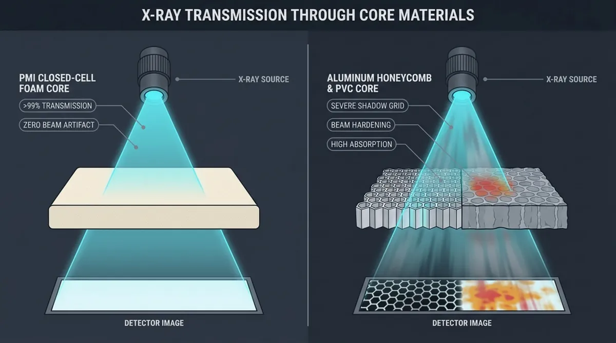
Polyethylene terephthalate (PET) and polyvinyl chloride (PVC) foams are widely used in industrial sandwich structures due to raw-material cost factors. However, PVC is unsuitable within an X-ray imaging path because it contains chlorine (atomic number 17). Chlorine interacts strongly with X-ray photons via the photoelectric effect, causing significant radiation attenuation, beam hardening, and severe imaging artifacts.
PET foam consists of carbon, hydrogen, and oxygen, but standard commercial grades possess larger, less uniform cellular structures than PMI. Extrusion foaming can produce density gradients, flow lines, and internal voids. In high-resolution mammography, these variations project visible artifacts onto the detector array, making PET unsuitable for primary imaging apertures.
PMI foam versus polyurethane foam
Rigid polyurethane (PU) foam is readily moldable but offers a lower specific modulus and lower shear strength than PMI. To match the mechanical stiffness of a 52 kg/m³ PMI core, an engineer must specify a significantly denser PU formulation, which increases component weight and radiation absorption.
Injected PU cores can also suffer from uneven density distribution and void formation during foaming. These inconsistencies cause non-uniform X-ray transmission. Additionally, PU exhibits lower thermal degradation thresholds, preventing its use in co-curing cycles alongside high-temperature prepregs in autoclaves.
PMI foam versus aluminum honeycomb
Aluminum honeycomb exhibits excellent stiffness along its cell axis, making it popular in primary aerospace structures. However, it cannot be used within a diagnostic beam path. Aluminum (atomic number 13) attenuates X-rays, and vertical cell walls project a visible hexagonal shadow grid onto digital image detectors.
Furthermore, honeycomb cores are structurally anisotropic. They provide high compressive strength along the cell direction but lack multi-directional shear strength across the plane. PMI foam provides isotropic mechanical support and maintains consistent radiolucency regardless of projection angles.
PMI foam versus solid thermoplastics
Solid polymers like polycarbonate, PEEK, and ABS are standard choices for injection-molded housings and transparent compression paddles. Polycarbonate paddles remain common in mammography because they give clinicians visual feedback during initial breast positioning.
Structurally, however, monolithic plastics require substantial wall thickness to limit flexure, significantly increasing weight. A monolithic plastic plate deflects considerably more than a PMI/CFRP sandwich panel of equal mass. Under continuous clinical loads, large solid thermoplastic plates can also undergo viscoelastic creep, resulting in permanent dimensional sag.
Choosing between minimum weight, cost, and structural performance
Core material selection balances structural requirements, radiolucent limits, and overall project costs. For non-structural housings located completely away from the radiation beam, general-purpose PET or reaction-injection-molded (RIM) polyurethane cores may suffice.
When a component resides within the imaging aperture, must satisfy tight deflection limits (such as the 1.0 cm MQSA compression limit), and requires high-temperature composite curing, PMI foam is the appropriate engineering choice. Its performance benefits offset raw-material cost differences by streamlining composite manufacturing and meeting clinical imaging standards.
What Are the Limitations and Risks of PMI Foam?
While PMI foam provides significant structural advantages, engineering teams must recognize its practical limitations. Specifying PMI without accounting for its processing characteristics, brittle behavior, and environmental sensitivities can lead to manufacturing scrap or premature part failure.
Designers must factor these variables into early mechanical analysis and manufacturing plans.
Higher material cost than general-purpose foams
PMI foam requires a complex manufacturing process involving controlled chemical casting and high-temperature thermal imidization. This makes PMI more expensive on a raw-material basis than common PVC, PET, or polyurethane cores.
For bulky external covers that carry no structural load and sit outside the imaging beam, specifying PMI without clear weight or temperature requirements can needlessly increase component cost. Engineers should target its use in assemblies where radiolucency, high strength-to-weight performance, and dimensional accuracy justify the material selection.
Potential damage from unsuitable adhesives or solvents
Fully cured PMI demonstrates good resistance to many industrial solvents and alcohols, but uncross-linked cell walls can be damaged by incompatible chemical systems during lamination. Uncured ester monomers, low-molecular-weight ketones like acetone, and specific amine curing agents present in certain room-temperature epoxies can soften or dissolve cell boundaries.
Chemical attack can collapse the foam surface cells, generating a dense, resin-rich boundary layer. This localized collapse undermines skin-to-core shear transfer and introduces an area of elevated X-ray attenuation that distorts diagnostic image data.
Reduced performance from incorrect machining or bonding
Due to its highly cross-linked chemical structure, PMI acts as a brittle-elastic polymer at ambient temperatures. Dull tooling, excessive feed rates, or incorrect tool geometries can fracture cell walls, induce subsurface micro-cracks, and chip edges. These machining defects become stress concentrations during cyclic mechanical loading.
Lamination errors present similar risks. Inadequate debulking or uneven vacuum pressure can trap air along the core-skin bond line. In mammography support plates, unbonded zones appear as visual artifacts on digital detectors and serve as initiation sites for fatigue delamination.
Need for application-specific fire and biocompatibility validation
PMI core materials do not automatically carry universal medical biocompatibility or flammability ratings. Standard industrial grades, such as Rohacell IG-F, are optimized for general lightweight composite structures and do not carry standalone ISO 10993 biological certification.
When an assembly requires specific flammability ratings, engineers must evaluate flame-retardant grades or ensure the external composite face sheets provide an adequate fire barrier. Assuming all PMI formulations possess identical medical-grade certifications will cause complications during regulatory compliance audits.
Why PMI foam should not be selected from density alone
Specifying a PMI core based strictly on nominal density (e.g., calling out a standard 52 kg/m³ core) is a common design oversight. While density correlates with compressive strength, it does not define cell size uniformity, resin uptake behavior, or shear fatigue limits.
Two foam grades sharing an identical density may exhibit distinctly different cell dimensions due to specific chemical formulations. A grade with larger cells absorbs more liquid resin during infusion or prepreg consolidation, increasing finished part weight and raising baseline X-ray attenuation. Specifications must clearly define cell size limits, resin absorption characteristics, and non-destructive testing requirements alongside nominal density.
What Manufacturing and Compliance Factors Should Medical Designers Check?
Transitioning an advanced composite medical device from prototype validation to serial production requires rigorous quality management. Material characteristics verified in development must be consistently maintained across production volumes.
Engineering teams must establish comprehensive quality verification procedures covering raw materials, core preparation, and finished laminated assemblies.
Material traceability and batch-to-batch consistency
Regulatory frameworks such as ISO 13485 and FDA 21 CFR Part 820 require manufacturers to maintain complete material traceability from chemical production to clinical deployment. Every PMI foam lot should be backed by a Manufacturer’s Certificate of Analysis (CoA) documenting core density, compressive strength, and thermal metrics.
Batch-to-batch consistency is essential for repeatable imaging calibration. Subtle fluctuations in base polymer density or residual reaction catalysts can alter the linear attenuation coefficient of a finished plate, which can disrupt the automated exposure control (AEC) calibration of a mammography system. Incoming inspection should verify sheet thickness, density tolerances, and purity.
Biocompatibility requirements for patient-contacting surfaces
While the PMI foam core remains fully encapsulated within the sandwich laminate, the finished medical assembly must satisfy ISO 10993-1 biocompatibility standards. If a compression assembly or patient support table contacts patient skin, external surfaces must be certified non-cytotoxic, non-irritating, and non-sensitizing.
The protective composite skins, surface gel coats, or polyurethane finishes must completely seal the inner core. The outer finish must resist chipping, peeling, or micro-cracking across years of continuous clinical handling, ensuring that patient fluids or topical lotions never penetrate the cellular core.
Cleaning, disinfectant, and sterilization compatibility
Medical hardware undergoes aggressive clinical disinfection daily. Design documentation must confirm material compatibility with typical hospital cleaning solutions, including accelerated hydrogen peroxide, glutaraldehyde, quaternary ammonium formulations, and 70% isopropyl alcohol.
Engineering evaluations must verify that these chemical agents do not craze exterior composite resins or breach perimeter seals. If the device requires low-temperature sterilization via ethylene oxide (EtO) or vaporized hydrogen peroxide (VHP), validation must confirm gas outgassing rates, aeration schedules, and structural cavity integrity under vacuum cycles.
Radiographic, mechanical, thermal, and environmental testing
Component qualification requires multiple validation testing protocols. Radiographic inspections, conducted via micro-CT or high-resolution flat-panel digital radiography, inspect completed sandwich panels for internal voids, resin rich pockets, cellular distortion, or foreign inclusions that could interfere with clinical interpretation.
Mechanical qualification involves static load and cyclic fatigue evaluations to verify compliance with deflection limits, ensuring total flexure remains under the 1.0 cm ceiling during clinical compression. Environmental testing must include hydrothermal aging. Environmental exposure studies show that combined heat and humidity (such as prolonged exposure to 80°C at elevated relative humidity) can reduce the compressive strength and stiffness of unsealed PMI panels by 30% to 50%. Finished assemblies must be tested post-aging to verify edge sealing performance.
Medical-device quality systems and supplier documentation
Device manufacturers sourcing structural foam cores or precision-machined sandwich components should partner with distributors operating under certified quality systems. Suppliers certified to ISO 9001 or AS9100 demonstrate established process control, tooling maintenance, and clean handling protocols.
Procurement specifications should mandate First Article Inspection (FAI) reports, non-destructive radiographic testing records, and formal change-control agreements. A documented change-control process ensures the material supplier cannot alter base polymer formulations, catalysts, or processing aids without prior written notification and validation by the medical device manufacturer.
How Should Engineers Specify PMI Foam for a Medical Application?
Translating the material benefits of PMI foam into a reliable, serial-production clinical device requires unambiguous engineering drawings and specifications. Vague drawing callouts, such as “PMI core, 52 kg/m³,” leave room for process variation that can result in quality defects.
Design teams should apply a structured specification methodology addressing radiological, mechanical, and environmental criteria.
Define the imaging path and allowable X-ray attenuation
Begin by establishing the maximum allowable X-ray attenuation for the structural panel. Specify the operational radiation spectrum, such as 25 to 35 kVp for mammography or 80 to 140 kVp for CT and general radiography. Establish an aluminum-equivalent ceiling-for example, requiring total sandwich transmission to exceed 98%, equivalent to less than 0.8 mm Al at the defined kilovoltage.
The specification must also set limits on attenuation variation across the active imaging area. A typical specification requires that localized radiation transmission vary by no more than ±0.5% to ±1.0% when evaluated with a calibrated diagnostic phantom, preventing internal core joints or surface filling compounds within the exposure window.
Set density, thickness, strength, and stiffness targets
Once radiological constraints are defined, calculate structural limits based on maximum clinical loading conditions. Clearly state the core density and allowable tolerance (e.g., 52 kg/m³ ± 5 kg/m³), compressive strength under ASTM D1621, and core shear modulus under ASTM C273.
Establish tight limits on core thickness and surface parallelism. Mammography detector platforms frequently require core thickness tolerances within ±0.05 mm or ±0.1 mm. Tight dimensional control ensures even bonding contact during composite curing, preventing the formation of resin-rich pockets that add weight and scatter X-ray photons.
Confirm operating temperature, humidity, and chemical exposure
Product specifications must clearly state ambient operating and storage limits. Typical operating ranges span 15°C to 35°C in clinical settings, while shipping and storage environments can fluctuate from -20°C to +60°C. Detail the relative humidity range and specify condensation resistance requirements.
List all approved hospital disinfectants and cleaning chemicals. Mandate that all exposed laminate edges, cutouts, and fastener counterbores be encapsulated with a certified, non-porous edge sealant to block moisture intrusion during daily clinical wipe-downs.
Specify surface skins, adhesives, inserts, and finish tolerances
A sandwich structure functions as an integrated system. Engineering drawings should detail the complete composite schedule, including carbon-fiber fabric type, fiber orientation, and prepreg resin content. A symmetric, quasi-isotropic layup (such as [0/45/90/-45]s) is commonly specified to prevent panel warpage during cooldown. Target resin content is typically set between 35% and 40% by weight to ensure proper fiber wet-out without excess resin pooling.
Detail all mechanical fastening interfaces. Specify the adhesive film system for core-to-skin bonding, the compressive modulus of structural potting compounds, and the specific thread depth, pull-out force, and torque limits for every embedded metallic or solid-plastic insert.
Validate prototypes under realistic clinical-use conditions
Finite element analysis (FEA) and analytical models must be validated using physical prototypes manufactured with production-intent tooling and curing cycles.
Validation testing should include proof testing at peak compression forces (e.g., 200 N), cyclic fatigue loading matching the full expected operational life (often exceeding 100,000 cycles), and radiographic verification using medical calibration phantoms. Test results must confirm zero measurable image degradation, cell damage, or permanent panel deflection over the simulated operating lifespan.
What Questions Should Buyers Ask PMI Foam Suppliers?
Procuring raw structural cores or machined kits for regulated medical devices requires detailed technical qualification. Engineering and procurement teams must confirm that a material partner can maintain tight quality controls rather than relying on generic product claims.
Targeted technical inquiries will help identify supply chain risks, manufacturing limitations, and process constraints before production begins.
Can the supplier provide material certificates and test data?
A qualified distributor serving medical manufacturers must furnish comprehensive documentation for every production lot. Essential questions include:
- Do you provide lot-traceable Certificates of Analysis (CoA) with every shipment, verifying density, compressive strength, and shear modulus against published specifications?
- Can you supply radiographic non-destructive testing (NDT) data confirming the foam core is free from metallic particles, chemical inclusions, and internal density streaks?
- Do you retain archive samples from each manufacturing lot, and what is your documented retention period under medical quality standards?
A supplier unable to provide comprehensive mechanical and radiological records creates regulatory compliance risks during notified-body or FDA audits.
What standard densities, thicknesses, and sheet sizes are available?
Confirming standard dimensional capabilities helps avoid unnecessary machining costs and long delivery lead times. Ask:
- What sheet formats do you keep in stock, and what maximum block dimensions are available prior to slicing?
- What are your standard horizontal cutting thicknesses, and can you supply thin sheets down to 1 mm while maintaining ±0.1 mm tolerances?
- Can you supply specialty grades, such as high-temperature variants like Rohacell XT, or provide custom densities for localized edge reinforcement?
Clarifying these processing metrics early prevents redesigns prompted by raw-material format limitations.
Can the foam be machined or supplied as a finished housing part?
Sourcing unmachined foam blocks requires maintaining internal 5-axis CNC routing equipment, dust extraction systems, and thermoforming tooling. For many medical device manufacturers, procuring precision-machined core components or pre-shaped kits is more cost-effective. Key questions include:
- Do you provide in-house 5-axis foam shape milling, horizontal slicing, and contouring services based on customer CAD models?
- What dust extraction and contamination-control procedures are utilized during foam machining to prevent surface debris from compromising subsequent composite bonding?
- Can you deliver ready-to-lay-up core kits with pre-beveled edges, thermoformed profiles, and close-tolerance insert pockets?
Procuring precision-milled cores directly from a foam-processing specialist reduces production waste, eliminates core prep labor, and keeps polymer dust out of cleanroom lamination environments.
Which adhesives, skins, and surface treatments are compatible?
Chemical incompatibility between core materials and processing resins can halt composite manufacturing. Inquire directly about processing parameters:
- Which epoxy, cyanate ester, or polyurethane resin systems and adhesive films are qualified for use with the selected PMI grade?
- What maximum curing temperature and pressure parameters do you recommend during autoclave or press-molding cycles to prevent cell distortion?
- Does the core require preparatory heat treatment or circulating-air drying prior to high-temperature resin infusion or prepreg consolidation?
Experienced technical partners supply detailed processing data sheets covering moisture drying schedules, thermal conditioning, and consolidation limits.
What quality controls apply to mammography and medical-device components?
The supplier’s quality infrastructure must match the high reliability demands of diagnostic radiology. Recommended questions include:
- Does your organization operate under established quality management systems such as ISO 9001 or AS9100?
- What inspection tools, such as optical CMM systems or digital radiography, verify the dimensional tolerances of precision-machined foam details?
- What formal customer notification procedure is executed prior to making changes to raw material suppliers, manufacturing equipment, or processing facilities?
Thorough supplier vetting ensures that device manufacturers capture the full mechanical, thermal, and radiological advantages of high-performance PMI cores throughout the operational lifespan of the medical equipment.
As diagnostic imaging trends toward photon-counting detectors, higher resolution, and faster examination workflows, structural composite components must satisfy tighter engineering tolerances. By delivering high stiffness-to-weight ratios, uniform cell structures, and minimal X-ray attenuation, PMI foam meets these clinical requirements. When paired with controlled composite lamination and rigorous quality verification, structural PMI foam serves as an enabling material for next-generation medical imaging hardware. If you are evaluating core materials for your medical composite application, contact Chem-Craft’s composite engineering team to discuss material selection and precision milling specifications.